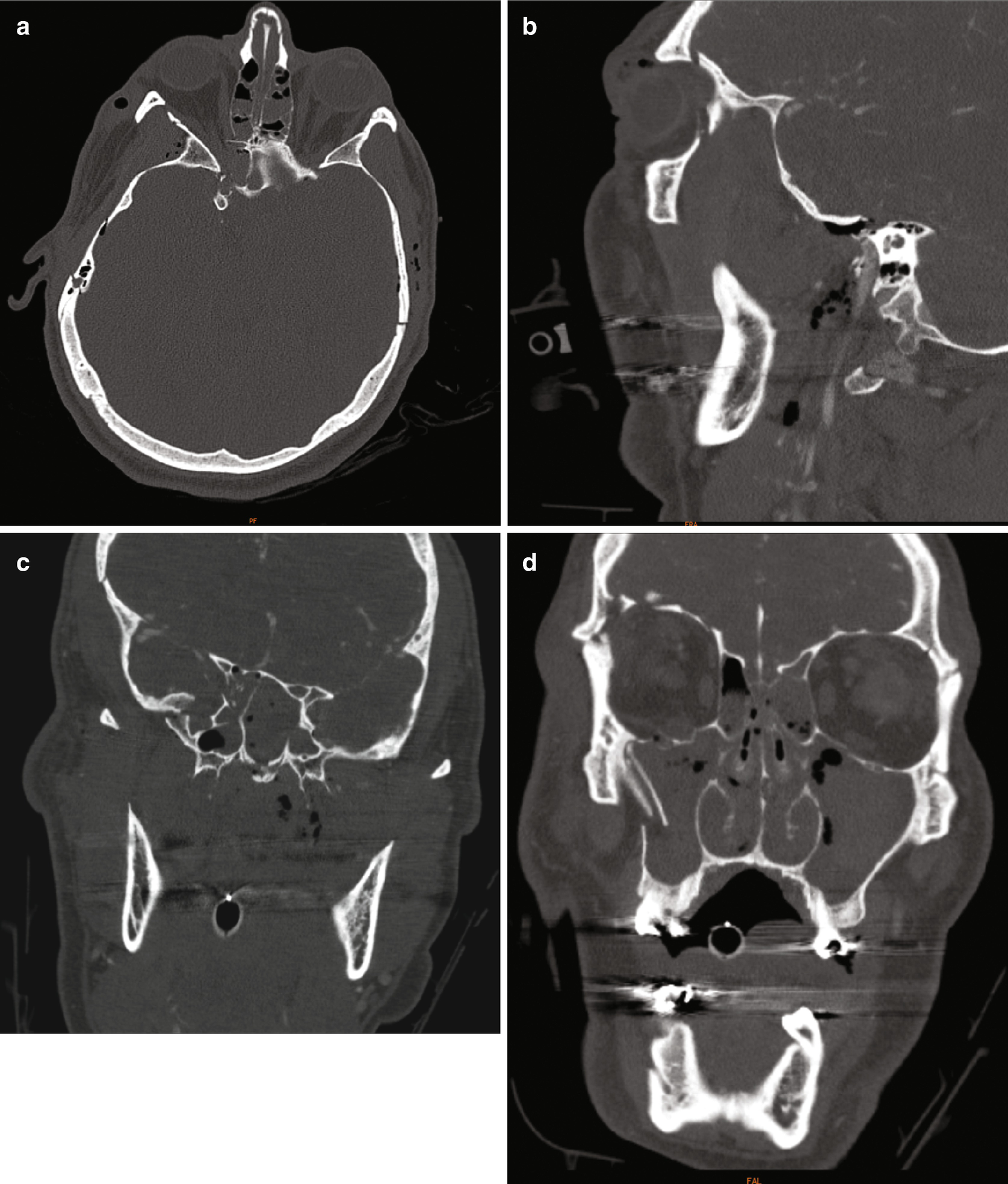
Ct Scan Orbital Floor Plate

History Of Prior Surgery Bone Defect Is Seen In The Right Cribriform Plate Contrast Leak And Accumulation Within The Righ Nasal Cavity Head And Neck History

Ct Scan Of Facial Bones Axial View A Showing Fracture Of Bilateral Download Scientific Diagram

Imaging The Face Radiology Key

Orbital Blowout Fracture Radiology Reference Article Radiopaedia Org
Https Www Neiltanna Com Assets Pdf Face 18 Pdf

Face And Neck Emergencies Radiology Key

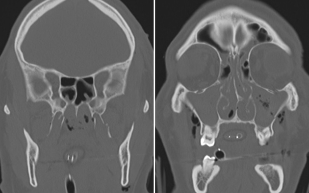
Coronal slice of a ct scan shows a non affected left orbit with normal anatomy of the transition zone.
Ct scan orbital floor plate. Universal 1 2 upper face module laser etched to aid in plate identification plate holding forcep plate holding forceps utilizes two pins to stabilize plate. Coronal slice of a postoperative ct scan taken after transconjunctival repair of the complete left medial orbital wall and orbital floor. Large fracture 50 of orbital floor on ct scan indicates that enophthalmos is likely to occur. Appropriate timing is based on the clinical exam and imaging.
The arrow indicates the buttress of the transition zone between medial orbital wall and orbital floor. These plates consist of implants that closely approximate the topographical anatomy of the human orbital floor and medial wall and are intended for use in a selective craniomaxillofacial trauma. Epidemiology the blowout fracture is t. We then cover the implant with our proven medpor biomaterial to minimize sharp edges even if the plate requires modification.
Computed tomography ct is the primary modality for assessing orbital soft tissue and bony injury in the emergency setting. Concomitant medial orbital wall fracture can increase risk of progressive enophthalmos. Plate borders medial wall orbital floor designed from ct scan data the three dimensional implants closely approximate the topographical anatomy of the hu man orbital floor and medial wall to provide accurate recon struction even after significant two wall fractures 5 6 preformed three dimensional shape. A coronal ct scan of a left maxillary mucocele eroding through the orbital floor and medial antral wall.
Ct scanning of the orbits is very quick which significantly reduces motion artifacts. We use ct scan data to design the titantium implants to approximate the anatomy of the orbital floor and medial wall. Silicone implants are 440 hu whereas in one study pmma implants were 135 hu 24. The following discussion assumes a volume ct technique using a multidetector scanner when referring to ct.
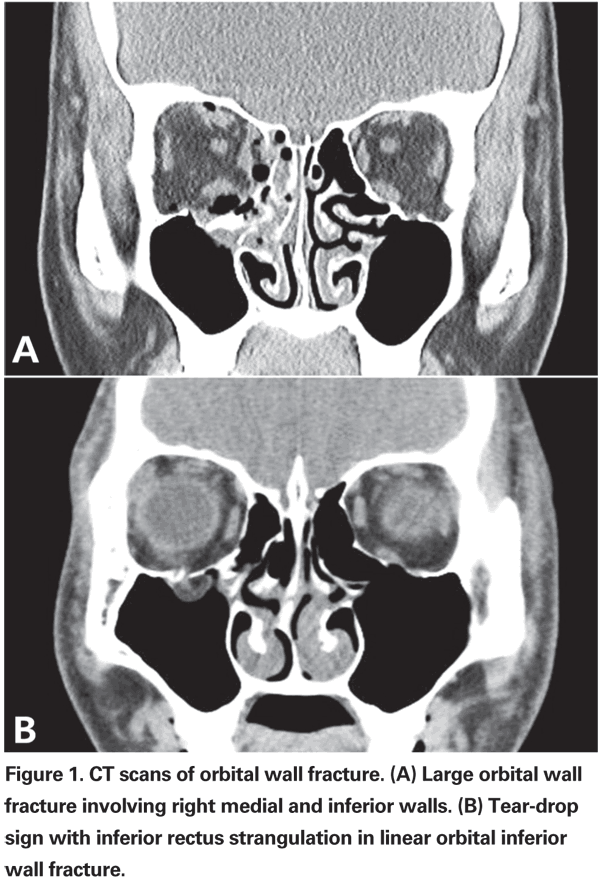
Materials such as silicone and pmma have been in use for over 30 years and are radiopaque fig 11. Orbital floor fracture repair might be indicated in this setting for small or medium sized defects. Orbital blowout fractures occur when there is a fracture of one of the walls of orbit but the orbital rim remains intact. This is typically caused by a direct blow to the central orbit from a fist or ball.
Functional endoscopic sinus surgery was performed to drain the maxillary mucocele and 50 ml of thick yellow mucus was expressed which was sent to pathology.
Diagnosis And Treatment Of Orbital Fractures
Https Www Zvitmedical Com Wp Content Uploads 2013 10 Doc 5 Pcl Permanent Versus Bioresorbable Implants In Orbital Floor Pdf

Intraoperative Imaging O Arm In Secondary Surgical Correction Of Post Traumatic Orbital Fractures Sciencedirect

Pin En Radiology

Intranasal Migration Of A 35 Year Old Orbital Plate Presenting As Unilateral Epiphora Sciencedirect
Open Reduction With Or Without Internal Fixation For Orbit Orbital Floor Fracture
Le Fort 3 Fractures Radiology Case Radiopaedia Org

Reconstruction Of Medial Wall Blowout Fracture Defect With A Combination Of Resorbable Meshed Plate And Cancellous Bone Allograft

Intraoperative Imaging Changes Management In Orbital Fracture Repair Journal Of Oral And Maxillofacial Surgery

Tips And Tricks In Surgical Management Of Maxillary Sinus Tumors Sciencedirect

Reconstruction Of A Complicated Orbital Depression Fracture With Medial Wall And Globe Repositioning In A Horse A Collaboration Across Disciplines And Specialties Mcmaster 2016 Veterinary Surgery Wiley Online Library
Orbital Reconstruction Springerlink
Le Fort Fracture Classification Radiology Reference Article Radiopaedia Org